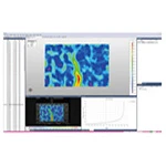
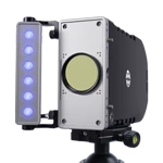
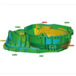
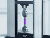
With the advancements in materials science, microelectromechanical systems (MEMS), and biomedical engineering, higher demands are being placed on the measurement of deformation and strain at the microscale. The Digital Image Correlation Micro-Strain Measurement System ( DIC HYPERLINK "https://www.xtop3d.com/products/xtdic-microxilie.html" \t "https://www.xtop3d.com/bconfig/_blank" ), with its advantages of non-contact, high precision, and full-field measurement, is becoming an important tool in laboratory and industrial testing. This article will provide an in-depth analysis of its working principle, key technical parameters, application areas, and purchasing recommendations to help researchers and engineers fully understand this advanced measurement technology.
I. What is a DIC microstrain measurement system?
Digital Image Correlation (DIC) is a non-contact measurement method based on image analysis . It calculates the strain distribution by continuously photographing the random speckle pattern on the surface of the object being measured and using algorithms to track the displacement of pixels.
Micro-DIC combines stereomicroscopy with DIC technology for deformation measurement at the micrometer or even nanometer scale, with resolution reaching the sub-micrometer level.
II. Composition of the Microscopic DIC Measurement System
A typical DIC microstrain measurement scheme consists of the following units:
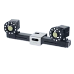
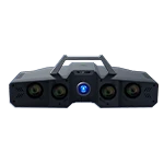
DIC Measurement System: Includes camera, light source, calibration plate and calibration device, speckle preparation kit and software;
Microscope: It adopts an optical magnification microscope with about 10x magnification and is compatible with two industrial cameras;
Temperature loading system: Supports programmable temperature control for both heating and cooling;
III. Detailed Explanation of Working Principle
The core of DIC lies in image correlation matching:
Speckle pattern preparation: Create a speckle pattern with random grayscale distribution on the surface to be tested (spraying, etching or natural texture).
Image acquisition: Continuous imaging of the sample surface under different loading conditions.
Subset matching: Select a small region (subset) in the reference image and find the best matching position in the deformed image.
Displacement field calculation: The displacement of each subset is determined by maximizing the correlation coefficient.
Strain field calculation: Differentiate the displacement field to obtain the strain distribution.
Based on this, the micro-DIC adds optical magnification and sub-pixel interpolation algorithms, enabling measurement accuracy to reach the nanometer level.
IV. Key Technical Indicators
XTOP3D microscopic DIC measurement technology:
Non-contact measurement technology;
XYZ 3D coordinate/displacement/strain full-field measurement; 1-10mm measurement field of view;
20µm highest strain measurement accuracy; 0.1µm warpage accuracy;
CTE determination; FEA comparison; maximum temperature range of ±190~600℃
V. Application Areas
Materials Science: Research on the mechanical behavior of metals, ceramics, and polymers at the microscale.
Microelectromechanical systems (MEMS): Deformation analysis of microstructures under stress, heat, and electricity.
Biomedicine: Measurement of the mechanical properties of cells and tissues, and microscopic strain analysis of biomaterials.
Electronic packaging: Reliability assessment of solder joints and chips under thermal cycling or mechanical loads.
Thin Films and Coatings: Measurement of stress distribution in thin films during bending and stretching processes.
VI. Recommendations for Selecting a Microscopic DIC Measurement System
Define the measurement requirements: scale, accuracy, and loading method.
Pay attention to camera and lens performance: resolution, noise level, and dynamic range.
Software features: Does it support 2D/3D DIC, thermo-coupling analysis, and automated processes?
After-sales service and technical support: training, maintenance, and upgrades.
Customization and scalability: To meet the testing needs of special operating conditions and potential future research directions.
FAQ
Q1: Can the micro-DIC system measure transparent samples?
A1: Transparent samples usually require surface treatment (such as spraying speckle) or the use of a special light source in reflective mode, otherwise it is difficult to obtain sufficient image contrast.
Q2: How to prepare the speckle pattern before measurement?
A2: Methods such as spray painting, laser etching, and nanoparticle deposition can be used. The key is that the speckle size should be adapted to the microscope resolution to avoid being too large or too small.
Q3: What are the advantages of micro DIC compared to traditional strain gauges?
A3: DIC provides full-field data rather than single-point measurement, does not damage the sample surface, and can adapt to complex deformation patterns, but the equipment cost and operation complexity are higher.
Q4: Is the system sensitive to environmental vibration?
A4: Very sensitive. It is recommended to use it on a vibration-damping platform or an air-bearing vibration isolation platform to ensure image stability.