The Challenge: Precision Control in Medical Injection Molding
Medical device cores, fluid flow-stop housings, and other medical injection-molded parts frequently feature complex free-form surfaces, deep internal cavities, sealing interfaces, and micro-coaxial structures. Factors such as overall surface uniformity, wall thickness distribution, concentricity, and injection molding warpage directly impact sealing performance, fluid control precision, and clinical reliability. Dimensional or surface deviations occurring across batches can easily lead to critical quality issues, including fluid leakage, loose assemblies, and uncontrolled flow rates.
Traditional measurement methods struggle to capture the complete 3D geometry of these components or quantify gradual surface deformations and hidden internal cavity defects. This significantly hinders efficiency in R&D mold tuning, mass production quality inspection, and regulatory compliance traceability.
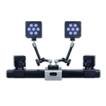
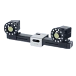
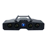
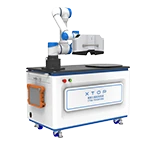
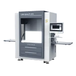
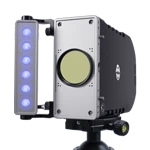
Leveraging non-contact full-field data acquisition, micron-level precision, and CAD-based full-field comparison and visualization capabilities, the XTOP3D XTOM stationary blue-light 3D scanner quantifies surface-wide dimensional deviations. It accurately identifies quality issues such as injection molding warpage, mold machining errors, and cavity wear, providing a comprehensive, digital, and traceable quality control solution for medical device manufacturing.
Advantages of Blue Light 3D Scanning Technology
1. Non-contact, Non-destructive Acquisition
Utilizing blue-light optical imaging for data acquisition ensures no scratching of polished mold cavities and prevents the deformation or compression of thin-walled medical plastic parts, making it ideal for the non-destructive testing of high-precision, high-cleanliness medical products.
2. Comprehensive Coverage of Internal Cavities and Micro-structures
It fully captures all internal and external surfaces, thin walls, and intricate features (such as steps) without measurement blind spots, accurately reconstructing full-field wall thickness and contour data.
3. Intuitive Quantification of Dimensional Deviations
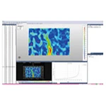
The scanned model is automatically aligned with the standard CAD model, intuitively displaying the distribution and magnitude of warpage, wear, and uneven thickness, thereby enabling the rapid identification of areas with quality issues.
4. Integrated Multi-dimensional Metrology
A single scan enables multi-dimensional inspection—covering surface deviations, cross-sectional thickness, concentricity, and hole position—and delivers comprehensive, quantified data in one step.
5. Digital Records for Compliance and Traceability
It automatically archives 3D models, deviation maps, cross-sectional inspection data, and standardized reports. This long-term storage supports longitudinal data comparison across batches and timeframes, meeting regulatory requirements for medical device quality compliance and traceability.
Typical Applications of Blue-Light 3D Scanning
The XTOP3D XTOM stationary blue-light 3D scanner is designed for the 3D inspection of various medical device components, such as polished metal cores, transparent or dark medical injection-molded parts, and complex thin-walled structures. By automatically aligning and comparing the scanned point cloud with the original CAD model, the system outputs full-field deviation maps, cross-sectional dimensions, geometric tolerances, and assembly gap data—providing a single-system solution for inspecting a wide range of typical medical device components. Case Study 1: 3D Inspection of a Precision Medical Mold Core
1. 3D Scanning Inspection Data
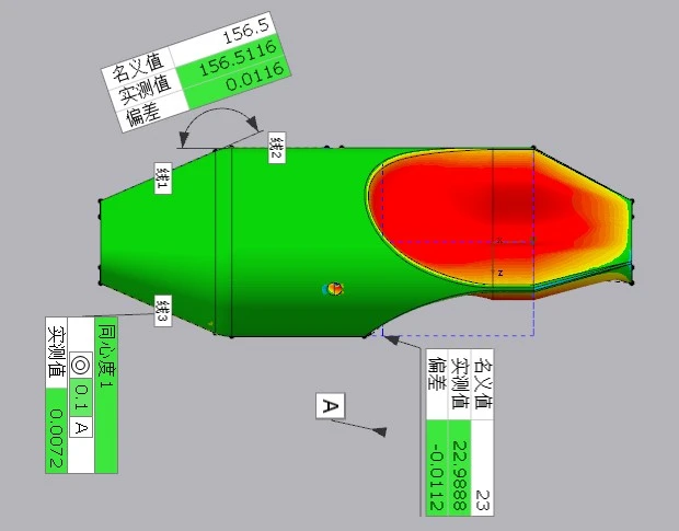
1) Geometric Dimensioning and Tolerancing (GD&T) Data: The nominal tolerance for core concentricity is extremely tight; measured values were 0.0072 mm and 0.0116 mm, with a maximum deviation from the nominal value of only 0.0112 mm. The geometric precision meets the standards for precision medical mold manufacturing.
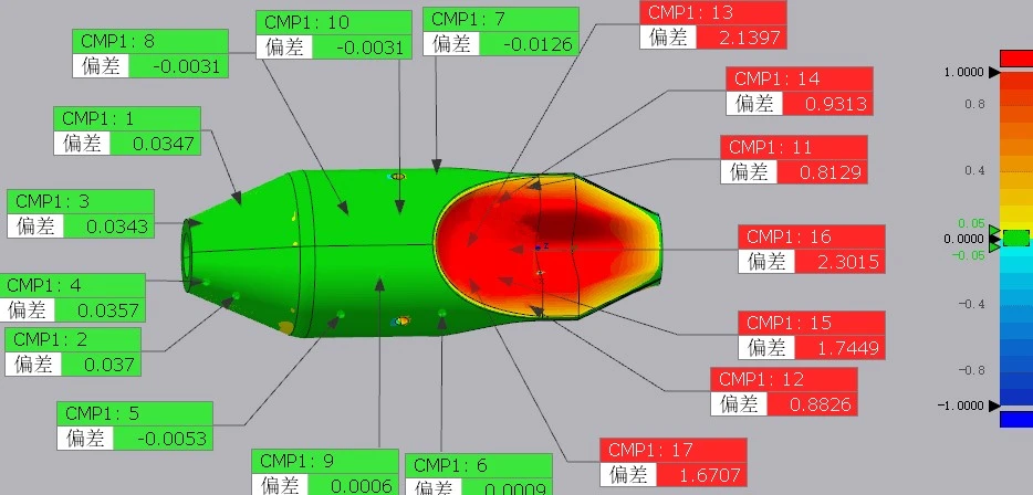
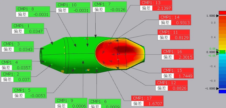
2) Full-Surface Deviation Color Map Data: The overall error range falls within ±0.01 mm, with deviations across the vast majority of the mold's forming surface controlled within a precision tolerance band of ±0.005 mm.
3) Comparison of Key Linear Dimensions: Differences between measured values and CAD nominal values for multiple hole diameters and cavity depths are at the micron level, with no significant dimensional drift.
2. Inspection Results
The overall machining precision of the medical mold core meets specifications. Core concentricity and cavity dimensional deviations are within the tolerances allowed for precision medical molds, ensuring that the molded product's sealing and coaxial assembly performance meet clinical standards.
The full-surface deviation map indicates only localized, minor machining errors at the micron level; there is no large-scale cavity deformation or polishing defects. No comprehensive mold rework is required, as optimization can be achieved through minor, localized polishing.
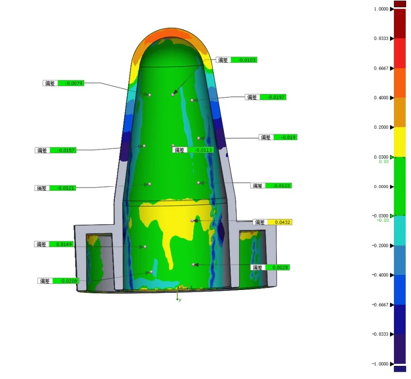
Case Study 2: 3D Inspection of an Injection-Molded Medical Flow-Stop Base
1. 3D Scanning Inspection Data
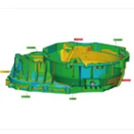
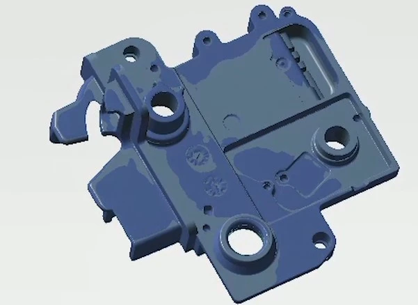
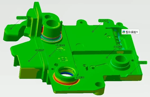
1) Global surface deviation range: Deviations across the majority of the part remained within the ±0.02 mm general tolerance for medical injection-molded components;
2) Sealing surface cross-section deviation data: A localized minimum deviation of -0.0295 mm was observed in the sealing contact area, indicating slight shrinkage and warpage from the injection molding process;
3) Assembly hole dimensional data: The difference between measured dimensions and CAD nominal values for multiple mounting reference holes was less than 0.015 mm; overall hole positional accuracy was within specifications.
2. Inspection Result Assessment
1) The overall dimensions of the flow-stop base comply with drawing requirements; however, localized slight shrinkage and warpage exist on the sealing surface. Although within tolerance limits, cumulative shrinkage during mass production could exacerbate these deviations, potentially leading to fluid leakage or unstable flow control over time;
2) A uniform, slight negative deviation was detected in the thin-walled section on the side of the base, caused by fluctuations in injection molding cooling parameters.
Case Study 3: 3D Inspection of a Thin-Walled Injection-Molded Medical Device Component
1. 3D Scanning Inspection Data
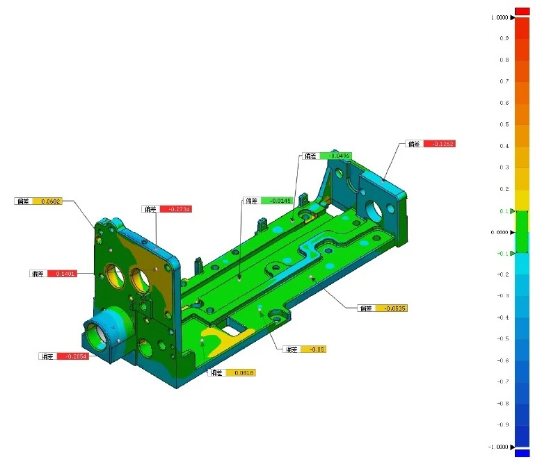
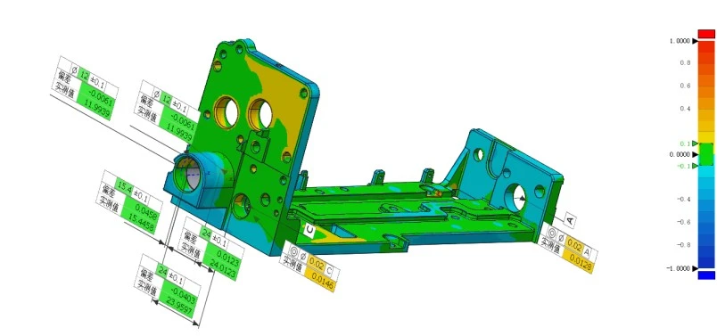
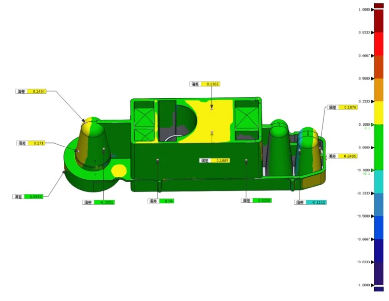
1) Global deviation color map range: Overall deviation ranges from -0.2794 mm to +0.1401 mm; deviations on the critical assembly surfaces of the main body fall within the ±0.1 mm standard tolerance zone.
2) Measured data for multiple sets of key linear dimensions: Deviations range from -0.0061 mm to +0.0458 mm.
3) Local warpage extreme values: The maximum negative deviation at the thin-walled edge is -0.2794 mm, indicating significant local warpage caused by spring-back during demolding.
2. Inspection Result Assessment
The assembly dimensions of the main body are within tolerance; however, the thin-walled edge area exhibits significant warpage that exceeds tolerance limits, which would result in excessive assembly gaps and sealing failure during mass assembly.
Multi-section thickness data reveals uneven wall thickness distribution across different regions, indicating room for optimizing injection molding holding pressure and cooling parameters.
Solution Summary
Precision molds for medical devices and thin-walled injection-molded parts demand rigorous tolerances regarding concentricity, sealing surfaces, wall thickness, and assembly clearances—factors that directly determine clinical safety. Contact-based inspection methods capture only discrete points, failing to provide comprehensive quality control for the entire surface, internal cavities, and thin walls; relying solely on such methods risks missing defects like extensive warping or deformation.
Powered by capabilities such as micron-level full-field scanning, global point cloud stitching, CAD model comparison, and quantitative cross-sectional analysis, the XTOP3D XTOM stationary blue-light 3D scanner rapidly identifies surface deviations and optimizes injection molding and mold manufacturing processes. It addresses key industry challenges—including concentricity control, warping and uneven wall thickness, the inability to measure internal cavity structures, and difficulties in quality traceability—thereby providing medical device manufacturers with a digital 3D quality inspection solution that meets industry compliance standards.