3D printing technology is widely used for prototype verification and the trial production of components with complex, curved surfaces. However, the printing process—influenced by factors such as layer thickness, temperature, material shrinkage, and the use of support structures—often results in defects in the finished parts, including localized deformation, dimensional deviations, and irregular surface textures. Traditional inspection methods typically rely on single-point sampling, while laser scanning often struggles to capture fine textures or intricate details on complex, curved surfaces. Consequently, for freeform shapes or uniquely sculpted components, it is difficult to rapidly identify all defective areas, which hinders the iterative optimization of printing parameters.
The objects for this inspection are 3D-printed components from a consumer-grade 3D printing brand, consisting of biomimetic animal models and test parts designed for evaluating geometric angles. The client’s primary challenge is that certain structural dimensions are inaccurate following the printing process. To address this, the client requires comprehensive 3D data of the finished parts. By comparing the scanned models against the original design CAD files, it becomes possible to precisely locate dimensional deviations and manufacturing defects, as well as quantify surface errors—thereby providing actionable insights for optimizing printing parameters and refining the original digital models.
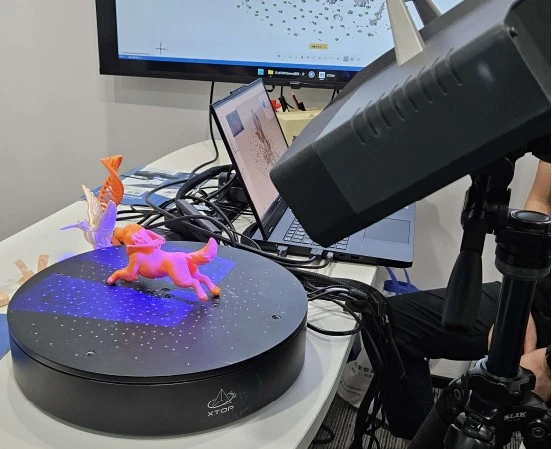
I. Limitations of Traditional Inspection and Laser Scanning Methods
1. Calipers and height gauges can only measure specific, localized dimensions; they cannot provide full coverage for complex curved surfaces, nor can they measure intricate corners, edges, or relief features.
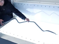
2. Coordinate Measuring Machine (CMM) inspection—which involves point-by-point measurement—is time-consuming. Because it only captures a limited number of discrete points, it is difficult to map the deformation distribution across large, complex surfaces.
3. Line-laser scanning: This method relies on a laser line to sweep across the workpiece surface. When dealing with features such as the fine layer lines characteristic of FDM printing, shallow relief, or subtle folds, the sampling density is often insufficient; this leads to a loss of detail and the "smoothing out" of layer lines. Furthermore, scanning complex surfaces can result in missing data—particularly on thin-walled sections or sharp corners—where the laser line may fail to register. In areas with abrupt surface transitions, the laser line is prone to reflections or "jump points" (erroneous data points). Capturing complete, accurate detail in these areas requires extensive adjustments to the scanning path, which significantly increases the time required.
4. Identifying defects solely through visual inspection fails to provide precise deviation values, which hinders effective process review and optimization.
II. Advantages of Blue-Light 3D Scanning in Capturing Fine Details
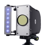
The XTOM blue-light 3D scanner utilizes area-based blue-light projection, allowing for the acquisition of a high-density 3D point cloud across an entire area in a single capture—eliminating the need for line-by-line scanning.
It is highly effective at capturing microscopic surface topography, including the intricate layer lines of 3D-printed parts, shallowly engraved characters, sharp tips, surface folds, and minute depressions or protrusions.
For complex, free-form surfaces with significant variations in contour, a single projection can cover a large area. Data loss is minimized—even in regions featuring sharp corners, thin walls, or complex transitions—without the need to repeatedly reposition the equipment or adjust the scanning path, thereby balancing high acquisition efficiency with exceptional detail reproduction.
This technology is particularly well-suited for assessing the surface quality of FDM-printed parts. Beyond measuring dimensional deviations, it can accurately replicate the microscopic textures resulting from the printing process, enabling the identification of issues such as irregular layer stacking or localized material accumulation.
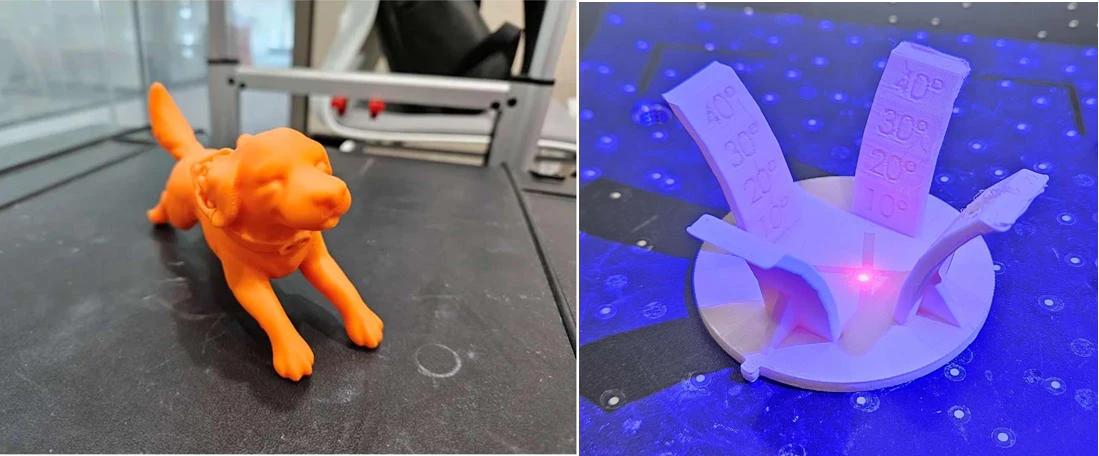
3D-printed thin-walled objects featuring biomimetic animal forms and relief-carved scales
III. Selection of Inspection Equipment
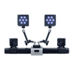
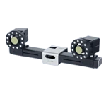
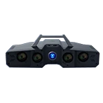
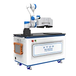
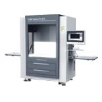
Given that 3D-printed parts often feature complex curved surfaces and intricate textural details, the XTOM-MATRIX-9M binocular blue-light 3D scanner was selected for inspection. When paired with an automated rotary table, it enables fully autonomous, 360-degree scanning and data acquisition of the workpiece.
Utilizing a structured-light (area-scanning) imaging approach, the high-resolution camera can accurately capture fine features—such as printing layer lines, sharp edges, and embossed text—thereby overcoming the limitations of laser scanning in capturing minute surface textures.
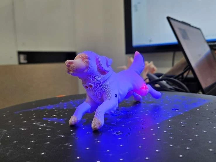
IV. Results of Detail Capture via Blue-Light 3D Scanning
Using the XTOM-MATRIX-9M blue-light 3D scanner, complete 3D mesh models were successfully generated for two types of printed parts: a biomimetic animal figure and a thin-walled component featuring embossed scales. Intricate structural details—such as printing layer lines, embossed characters, curved surface transitions, and sharp, thin-walled edges—were all faithfully reproduced.
4.1. Scanning Results for the Biomimetic Animal Figure
The 3D model of the biomimetic animal figure, obtained through blue-light 3D scanning, allows for the clear identification of several manufacturing defects, including deformations, material accumulation, and missing edges or corners on the ears, tail, paws, and the sides of the body.
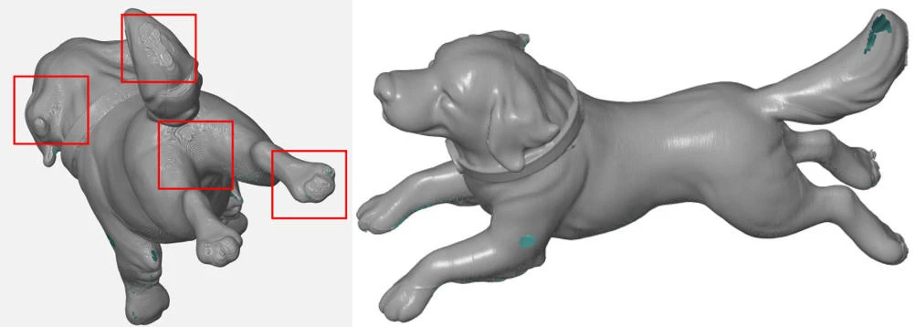
Bionic animal prototype defect marking & overall scanning 3D mesh
The scan details clearly reproduce microscopic features such as FDM print layer lines, the tips of the claws, and the small holes in the collar:
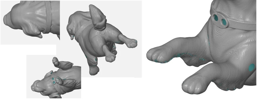
Scan results for specific body details & paw details
4.2 Test Results for the Angular Scale Specimen
The 3D-printed specimen featuring embossed scales contains several thin-walled, bent structures. The scanned mesh accurately reproduces the geometry of the base, the upright plate, and the embossed characters. The scan results clearly reveal printing defects, including warping and deformation in the thin-walled sections, edge imperfections on the embossed characters, and localized material accumulation.
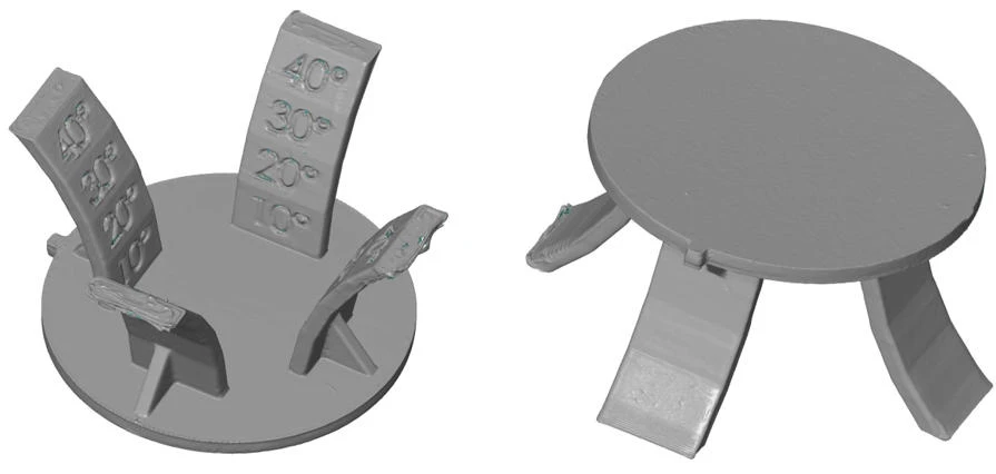
Overall 3D data model from sample scanning
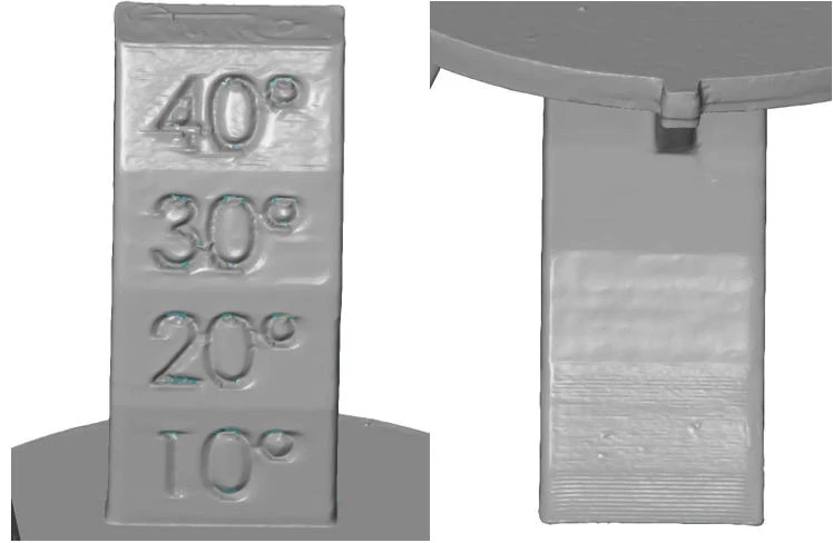
Scanning details of scale markings & thin-walled sections on the sample part
V. Quantitative Deviation Analysis
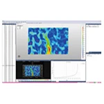
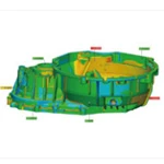
By scanning the 3D-printed prototypes and aligning the resulting meshes with the original CAD design models, it is possible to generate color-coded deviation heatmaps and detailed data tables. This enables both the qualitative identification of manufacturing defects and their precise quantitative assessment.
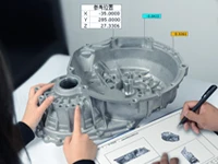
Users can access nominal dimensions, actual measured dimensions, and specific deviation values for any given point. This facilitates the rapid pinpointing of problematic areas, allowing for the review of printing parameters, modification of original CAD models, and iterative refinement of the manufacturing process.
VI. Case Study Summary
The XTOM-MATRIX-9M blue-light 3D scanner, when paired with an automated rotary table, is well-suited for analyzing dimensional defects in 3D-printed components with complex, curved surfaces. Through the 3D scanning of representative test objects—such as biomimetic sculptures and thin-walled carved pieces—the system’s area-based (structured-light) capture technology ensures the high-fidelity preservation of fine details, including FDM layer lines and embossed characters.
Non-contact blue-light 3D scanning allows for the complete digital reconstruction of a printed part's surface. By performing a full-field comparison between the actual finished part and the original CAD design, users can intuitively locate printing defects—such as deformation, shrinkage, or material accumulation—while simultaneously generating quantitative deviation data. This blue-light 3D scanning solution provides essential digital data to support the optimization of 3D printing parameters, model refinement, and quality control, ultimately enhancing the efficiency of quality verification for prototypes.